Faulty Molecular Transport Process May Lead to Movement Nerve Cell Death, Study Reports


A faulty molecular transport process could explain how loss of a protein leads to the death of movement nerve cells that is seen in spinal muscular atrophy, a study reports.
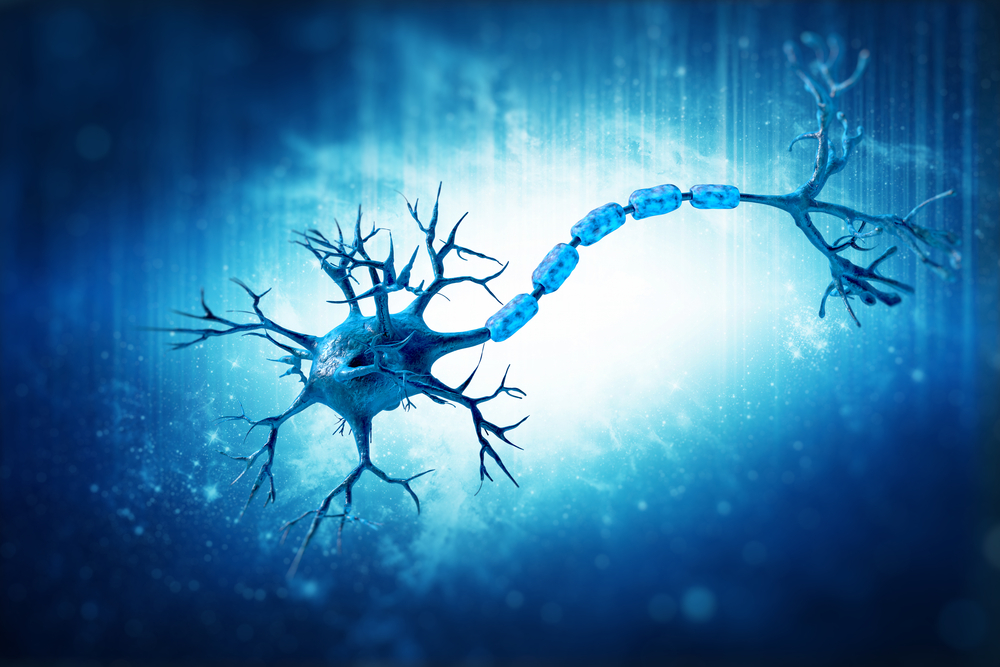
The key may be movement nerve cell projections’ inability to grow, Ohio State University researchers said. The projections, known as axons, pass on signals between nerves and muscles.
A team at the university’s Wexner Medical Center discovered that axon growth requires survival motor neuron protein interacting with another protein, HuD. The two likely transport messenger RNA molecules needed for axon development. Lack of survival motor neuron protein, or SMN, prevents the interaction from working properly.
Increasing HuD in zebrafish with no SMN improved both nerve cell development and the movement difficulties seen in SMA, according to the study, published in The Journal of Neuroscience. The team titled the article “HuD and the Survival Motor Neuron protein interact in motoneurons and are essential for motoneuron development, function and mRNA regulation.”
It’s likely that HuD is not the only factor interacting with SMN during movement nerve cell development, researchers said. They said work is needed to identify the others.
Although scientists know that mutations of the SMN gene cause SMA, they have yet to discover how the mutations cause movement nerve cells to die. Earlier studies indicated that SMN protein binds to what scientists call RNA-binding proteins in nerve cells, including HuD.
The Ohio State team decided to see if SMN’s interaction with HuD could influence axon growth.
When they looked at zebrafish movement nerve cells, they discovered that their suspicion was correct. They also discovered that the two proteins interact only around the time axons are developing.
Another finding was that the levels of the two proteins were tightly linked: When SMN levels were low, HuD levels were, too. Again, the team only saw this early in the fish’s development.
When the researchers genetically engineered zebrafish to lack HuD, they noticed that the fish had similar movement nerve cell abnormalities as fish models of SMA. The animals also had similar movement defects, suggesting the two proteins were involved in both processes.
An additional experiment supported this idea. When the team manipulated fish that lacked SMN protein to increase their production of HuD, the animals developed milder movement nerve cell abnormalities and fewer movement problems.
Because lack of both SMN and HuD led to lower levels of a messenger RNA molecule crucial to axon growth, the team concluded that the two proteins interact to transport the messenger RNA. Messenger RNA are intermediates in the process of producing a protein from a gene.
Research suggests that RNA-binding proteins like HuD may transport the molecule to another part of the axon, preventing the molecule from turning into a protein too soon. Lack of RNA-binding proteins can lead to messenger RNA deterioration, the study suggests.
The research failed to help the team understand why increasing HuD in fish that lacked SMN could improve, but not completely reverse, movement nerve cell abnormalities. One explanation, they said, could be that still other RNA-binding proteins are involved in the process.
Following up the study could explain how lack of SMN causes movement nerve cell death, the researchers said.