Muscle Thickness Decreases Over Time in SMA Type 1 as Measured by Ultrasound


Children with spinal muscular atrophy (SMA) have weaker muscles than children who are not affected by muscle myopathies and new results from a study conducted at Washington University School of Medicine in St. Louis, Missouri, indicate this muscle weakness may be due to differences in muscle thickness. Researchers used ultrasound to measure muscle thickness in the arms and legs of three children with SMA and found negative changes over time.
As described in “Quantitative Muscle Ultrasound Measures Rapid Declines Over Time in Children with SMA Type 1,” which was published in Journal of the Neurological Sciences, children with SMA type 2 and 3 do not show changes in muscle size over time. However, it was unknown if muscle size changes over time in children with the more severe SMA type 1. In order to answer this important question, a research team led by Dr. Kay W. Ng identified three infants–aged one, six, and eleven months–who had SMA type 1 and measured their muscle thickness using ultrasound. Measurements were repeated two or four months later.
Although at baseline the children showed normal muscle thickness, except for the quadriceps (thigh) muscle in the oldest child, at the later time point muscle thickness decreased. All three children showed lower than normal quadriceps muscle thickness. Negative changes were also noted in the biceps of two children and the anterior forearm of one child, but the tibialis anterior (shin) muscles were unchanged in all three. This indicates that not all muscles are affected equally throughout time by SMA type 1–muscles closer to the body were more affected than those further from the body.
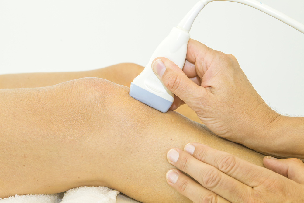
The researchers were especially interested in using ultrasound for their study because it is a relatively simple and less painful technique to measure muscle thickness and function in children. Other researchers have used magnetic resonance imaging (MRI) or compound muscle action potential (CMAP) testing, but these techniques require high levels of training for hospital personnel and may be painful for children. Ultrasound is different in that it uses pulses of sound to create an image that can then be analyzed for thickness and cross sectional area with accuracy similar to that of MRI.
Although it has been shown that muscle size does not change in children with SMA types 2 and 3, this new study suggests that ultrasound may be an important tool for clinicians who see SMA patients, since it can detect changes in muscle size in children with the more severe SMA type 1.