Magnetic Rods May Be Alternative to Correct Scoliosis in Children With SMA, Study Suggests

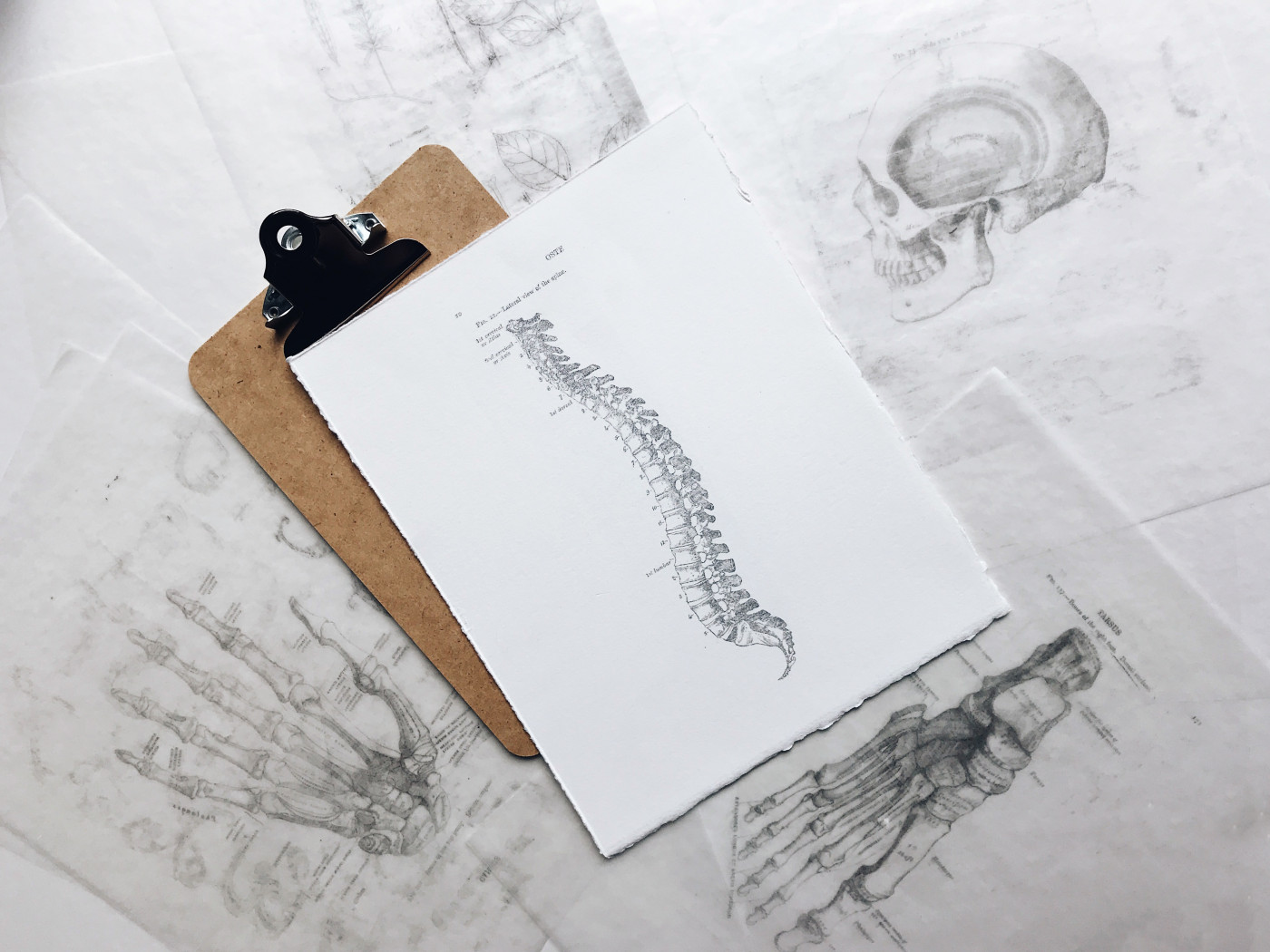
Photo by Joyce McCown on Unsplash
Spine surgery using magnetically controlled growing rods (MCGR) may be an option to correct spinal deformity, or scoliosis, in young children with spinal muscular atrophy (SMA) and avoid the need for repeated surgeries as children’s spine grows, according to a recent study.
The study, “Early Results of a Management Algorithm for Collapsing Spine Deformity in Young Children (Below 10-Year Old) With Spinal Muscular Atrophy Type II,” was published in the Journal of Pediatric Orthopaedics.
Scoliosis, an abnormal and progressive curvature of the spine, is a common health problem faced by SMA patients, and can worsen their breathing capacity and further limit their mobility.
Its severity is directly linked to SMA type — patients with type 1 or type 2 disease are likely to develop scoliosis, as are about 50% of individuals with SMA type 3.
The most common treatment for scoliosis caused by SMA is surgery, with early spine fusion the past treatment of choice. This strategy involves the surgical joining, or fusing, of the small bones of the spine, or vertebrae, but is inadequate for growing children as it cannot accompany the growth of the spine.
Ask questions and share your knowledge of Spinal Muscular Atrophy in our forums.
Over the past decade, there have been several advances toward more growth-friendly procedures, such as implanting growing rods, or expandable titanium ribs, on each side of the spine. The rods can be further lengthened with subsequent surgeries as the child grows.
More recently, a new approach — called magnetically controlled growing rods (MCGR) — was developed to bypass this need for repeated surgeries. This provides the critical advantage of minimizing surgery-related risks such as infections and pulmonary complications, as well as lowering costs and improving patients’ quality of life.
After being surgically implanted on each side of the spine, MCGR rods can be lengthened externally, in a non-invasive manner, using a remote control placed over the patient’s back.
Each rod has a magnet inside that rotates with the use of this external controller to lengthen or shorten as needed. Rod adjustment can be made during outpatient visits and causes little or no pain.
Spine-based MCGR has given encouraging results in groups of children with scoliosis, but no study had looked exclusively at the outcomes in children with SMA.
Researchers developed a management approach for using MCGR specifically in young children with SMA and report their early results in 11 patients with SMA type 2, ages 6 to 10, treated at the Cankaya Hospital in Turkey between 2014 and 2017. The mean follow-up time was 37 months.
All children had early-onset scoliosis, which appeared before age 10, and was characterized by progressive C-shaped spine deformity marked by an asymmetrical pelvis falling to one side, or pelvic obliquity.
Magnetic rods were surgically implanted on both sides of the spine in eight patients. In three patients, the magnetic rods were placed only on the concave side (curving in) of the spine together with a gliding rod on the convex side (curving out) because their spinal curves were too rigid and because the curve’s tip was outside a target zone.
Instruments were placed along the spine down to the pelvis in nine patients, while in two of the patients, the implants did not reach the pelvis and were placed only up to the lumbar zone, or L3 vertebra.
MCGR corrected spine curvature from an average of 81.8 degrees before surgery to 29 degrees after the procedure. On a follow-up visit at 35 months after surgery, the patients’ spinal curve was at had an average of 26 degrees.
The procedure also eased pelvic obliquity, which dropped from a mean of 20.9 degrees before the procedure to 4.9 degrees post-surgery, and remained at 6.5 degrees at the last follow-up.
As the children grew, the magnetic rods were progressively lengthened at outpatient visits. During follow-up, spinal height — between T1 and S1 vertebrae — increased from a mean of 329 mm after surgery to 356 mm, corresponding to a growth of 8.9 mm per year.
No neurologic problems, infections, or implant-related complications were noted. Additional deformities occurred in the two patients not receiving any pelvic implant, and these patients either underwent or were scheduled to undergo revision surgery to extend the implants to the pelvis.
“Short-term results indicate that MCGR may be a good option in SMA-associated collapsing spine deformity to reduce the burden of repetitive lengthening procedures,” the researchers wrote.
They recommended implanting instruments until the pelvis in all children, to prevent additional deformities. For those who have rigid and large spine curves, “single MCGR combined with a convex gliding rod may be a valuable option,” they said.
They also recommended further evaluation in longer-term studies to better understand the impact on pulmonary function and overall health.


