High-resolution MRI Scans of Spine May Offer Way to Detect and Monitor SMA, Case Study Suggests
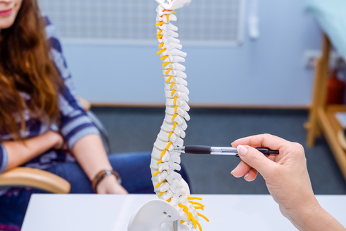


High-resolution magnetic resonance imaging (MRI) of the spinal cord may be a non-invasive way of monitoring spinal muscular atrophy (SMA) disease progression and responses to treatment, a case report suggests.
The technique helped clinicians to diagnose a toddler with SMA type 2 after it revealed signs of ventral nerve atrophy in his lower back.
The case report, “Lumbosacral ventral spinal nerve root atrophy identified on MRI in a case of spinal muscular atrophy type II,” was published in Clinical Imaging.
SMA is characterized by the gradual loss of motor neurons — the nerve cells responsible for controlling voluntary muscles — in the spinal cord, leading to muscle weakness and wasting. It is divided into subtypes (ranging from type 0 to type 4) depending on the age of disease onset and symptom severity.
Ventral nerve root atrophy and anterior horn cell degeneration is considered hallmark features of the disease, but often found during post-mortem examinations of patients. (Ventral nerve roots enable motor neurons that control muscles and glands to exit the spinal cord, along with other neural signals from the central nervous system; anterior horn cells are specific motor neurons located in the spinal cord that affect skeletal muscles.)
“Despite the prominence of these pathological features, previous studies of spinal imaging in SMA have reported normal findings in the lumbosacral spine [lower back] or did not inspect the lumbosacral spine,” the researchers wrote, noting that advanced and 3-D high-resolution scans are likely necessary for SMA patients.
Interested in SMA research? Check out our forums and join the conversation!
The boy in this case study began to have difficulties with latching during breastfeeding and bottle feeding when he was 2 months old. Nevertheless, he hit most developmental milestones, such as sitting, crawling and standing by himself, during his first year of life.
But by 19 months old, physicians noticed he had poor muscle tone, especially in his legs and feet, and delays in developing age-relevant motor skills. An MRI brain scan showed no abnormalities. But signs of ventral nerve atrophy, compared to an age-matched healthy infant, were found in scans of his lower spinal cord.
Genetic tests were then performed and confirmed an SMA type 2 diagnosis. The boy had been given four doses of Spinraza (nusinersen) by Biogen, the only approved treatment for SMA type 1, 2 and 3, administered through a spine injection, before this case study ended.
At age 29 months, he was able to stand and to walk without the help of a walker. His parents also reported significant improvements in his strength and mobility since treatment began.
“This case illustrates that spine MRI may be helpful for evaluation of ventral nerve root atrophy in SMA. The next step would be to analyze additional spine MRI data, both retrospectively and prospectively, to determine how consistently these findings are present in SMA, and to explore the potential use of advanced high-resolution spine MRI as a non-invasive biomarker for monitoring disease progression and responses to novel therapies,” the researchers concluded.