Children with SMA at Risk of Weak Bones and Fractures, Study Finds
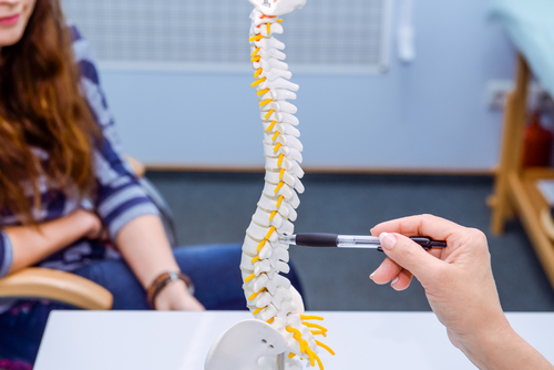


Young children with spinal muscular atrophy (SMA) types 2 and 3 are at risk of severe bone fragility and can carry fractures that go unnoticed, a study found.
The results, “Evolution of bone mineral density, bone metabolism and fragility fractures in Spinal Muscular Atrophy (SMA) types 2 and 3,” were published in the journal Neuromuscular Disorders.
With the recent breakthrough advances in SMA treatment, “there is a strong need to fill the gap in the knowledge on the effects of the disease on different organs and systems, including bone, and the possible efficacy of therapies on the resultant comorbidities,” the researchers said.
Low mobility typically results in bone mass, density, and strength deficiencies, causing weak bones. For this reason, children with low muscular mass and reduced mobility, including those with SMA, are at high risk of fractures.
Additionally, preclinical data suggest that low levels of survival motor neuron (SMN) protein — the underlying cause of SMA — may also be directly involved in bone mineralization problems. However, to date, there is limited knowledge on the extent and features of bone involvement and its correlation with motor abilities in SMA.
Researchers at Istituto Neurologico Carlo Besta, Italy, and the Dubowitz Neuromuscular Centre, University College London (UCL), in the U.K., investigated the evolution of bone metabolism, bone mineral density (BMD), and fractures, along with their relationship with age and motor function in 32 children with SMA types 2 and 3 (mean age 3.3 years). Of these, 27 had SMA type 2 and five had SMA type 3 (four could walk independently).
The researchers had previously examined most of these children (29) and found that they had low levels of vitamin D and high levels of parathyroid hormone (PTH), two messenger molecules fundamental to regulating bone calcium. They also noted signs of increased bone breakdown and undiagnosed vertebral fractures.
Now, children underwent evaluations for diet, bone metabolism (laboratory tests), bone mineral density (bone density scan, or DXA), vertebral fractures (spine X-rays), and motor function (measured by the Hammersmith Functional Motor Scale Expanded (HFMSE) and the Upper Limb Module (ULM), at the study’s start and at 18 months of follow-up. Twelve children had an additional follow-up evaluation after 36 months.
Importantly, none of the children had received Spinraza (nusinersen) or any other investigational treatment for SMA.
At study start, all children began taking calcifediol (a form of vitamin D) and dietary calcium intake was adjusted according to recommendations.
The data revealed that low levels of vitamin D and asymptomatic (without symptoms) vertebral fractures were more common at the study start, highlighting the importance of correcting vitamin D deficiency to control PTH levels and avoid excess bone deterioration.
In the first evaluation, 10 children (31.2%) had low vitamin D levels and three had seven undiagnosed spinal fractures. At follow-up (18 months), six children (18.8.%) had low vitamin D levels and none carried vertebral fractures.
At the beginning of the study, four patients had a history of peripheral fractures in the limbs, while during the study five others developed similar types of fractures. Most were probably due to low- impact trauma and are likely a consequence of serious bone fragility, the researchers noted.
Markers of bone breakdown, or resorption — the process by which osteoclasts (bone cells) destroy bone tissue, releasing calcium into the bloodstream — were higher than normal at all visits, suggesting an excess of bone destruction.
Consistent with elevated bone destruction, bone mineral composition and density worsened over time, compared with study start. In particular, the percentage of patients with low spine mineral density reached 75% at 36 months.
No correlation was found between HFMSE motor scores and bone mineral content or density, but the researchers stress that higher values of spinal bone density at follow-up were associated with higher mobility scores at study start.
This points out that physically active children with SMA 2 and 3 may be more protected from osteopenia and osteoporosis (low bone mineral density), thus confirming the role of activity and muscle strength on bone gain in this young population.
Bone formation markers were within the expected range, or only mildly increased, “suggesting that bone formation was essentially adequate for age,” the researchers noted.
These findings suggest that “even young children with SMA are at risk of severe bone fragility,” the researchers said. “Our 36-month study confirms that fragility fractures are relatively frequent in children with SMA, especially in type 2.”
A better understanding of the molecular mechanisms behind bone metabolism alterations in SMA “could help to identify novel therapeutic targets and to establish better guidelines for the management of bone fragility in SMA.”
Long-term follow-up of patients treated with Spinraza, Zolgensma (onasemnogene abeparvovec-xioior) treatments currently under development “will provide further evidence on the possibility to counteract and/or recover bone abnormalities in these patients,” the researchers concluded.