Early nerve-muscle connection flaws set stage for SMA damage
Failure to mature soon after birth creates 'functional fragility' for NMJ
Written by |
- In SMA, the neuromuscular junction (NMJ) fails to mature normally after birth, making it vulnerable to damage.
- This immaturity leads to reduced neurotransmitter release, muscle weakness, and quicker fatigue, preceding motor neuron loss.
- Understanding NMJ maturation could provide biomarkers for SMA progression and treatment effectiveness.
A review study found that the neuromuscular junction (NMJ), the site where nerves connect to the muscles they control, fails to mature normally soon after birth in people with spinal muscular atrophy (SMA), leaving it structurally underdeveloped and functionally vulnerable before the loss of nerve cells that facilitate movement.
“We propose that delayed and incomplete postnatal maturation of the NMJ in SMA creates a state of functional fragility that precedes and predisposes [nerves] to later destabilisation and degeneration,” the researchers wrote.
The study, “Immaturity of the neuromuscular junction in spinal muscular atrophy mouse models,” was published in Frontiers in Cellular Neuroscience.
SMA is caused by a lack of SMN, a protein essential for the function of motor neurons. Without enough SMN, motor neurons degenerate, leading to SMA symptoms of muscle weakness and atrophy (shrinkage).
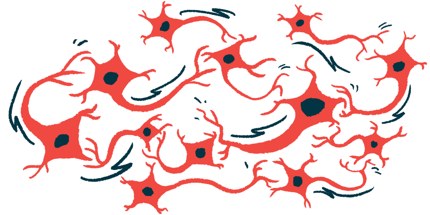
Emerging evidence suggests that, before overt motor neuron loss, there are signs of dysfunction at the NMJ, a highly specialized synapse where motor neurons communicate with muscle fibers to initiate muscle contraction. Specifically, electrical signals from the nerve fiber trigger the release of acetylcholine, a signaling molecule (neurotransmitter), which crosses the synapse and binds to receptors on muscle cells, triggering a muscle contraction.

Rapid development usually occurs soon after birth
A team led by researchers at the University of Seville in Spain conducted a review of published studies investigating the structural, molecular, and functional aspects of the NMJ in SMA, using mouse models, human tissue, and clinical data.
The NMJ typically undergoes rapid development soon after birth. As muscles grow, the nerve terminal (the end of the nerve that ends at the synapse) adapts. The number of acetylcholine release sites (active zones) increases, and the pool of synaptic vesicles (tiny sacs that store acetylcholine) expands. This allows more neurotransmitters to be released with each nerve signal.
In SMA, this developmental process is delayed and incomplete, with nerve terminals retaining features typically seen during early development. They have fewer active zones and smaller vesicle pools. Some muscles appear more affected than others.
Studies of NMJ structure show that vesicle clusters in SMA remain small and scattered, rather than becoming larger and more organized. The cytoskeleton, the internal support system of the nerve terminal, shows immature, disorganized features typically seen only early in life.
Changes also occur on the muscle side of the NMJ. Normally, the acetylcholine receptor switches from an embryonic form to an adult form. In SMA, this process is delayed, resulting in smaller, simpler endplates, the regions of muscle membrane that receive nerve signals. Muscle fibers are also smaller and mature more slowly.
At the molecular level, low SMN disrupts the handling of certain messenger RNAs (mRNA) in motor nerves. mRNAs serve as templates for the production of proteins, including those that build and maintain the NMJ. When this is impaired, fewer of these proteins are produced, which affects the formation of release sites, vesicle movement, and the organization of the nerve terminal. These problems appear early, before visible damage to the motor neuron.
Functionally, these structural and molecular changes reduce the amount of neurotransmitter released. In SMA, the number of vesicles released with each nerve signal can be reduced by up to 55% in more affected muscles during early development. Although release improves as the NMJ develops with age, it does not reach normal levels before the disease progresses.
The NMJ in SMA also has reduced capacity to sustain repeated activity. Normally, synapses can maintain output during ongoing use, but in SMA, they fatigue more quickly. This reflects a reduced synaptic reserve, a distinct group of vesicles that rapidly replenish acetylcholine. At the whole-muscle level, this defect contributes to smaller electrical responses and fewer functional motor units early in life.
Some of the proteins that help control neurotransmitter release are also altered. For example, there is a delayed switch between two forms of a calcium-sensing protein, with the more efficient adult form appearing at lower levels. Other proteins involved in preparing vesicles for release are also reduced or mispositioned.
As SMA progresses, additional problems develop. Transport of molecules within the nerve fiber becomes less efficient, proteins can abnormally accumulate, and mitochondria, which provide energy to the cell, do not function properly.
“These findings have important implications for understanding disease progression and therapeutic intervention,” the team wrote. “Parameters that reflect NMJ maturation … may serve as sensitive biomarkers of disease state and treatment efficacy, complementing traditional measures of motor neuron survival or muscle strength.”