
New Glia Cell Markers May Provide Insight Into Neuromuscular Diseases, Study Says
Written by |
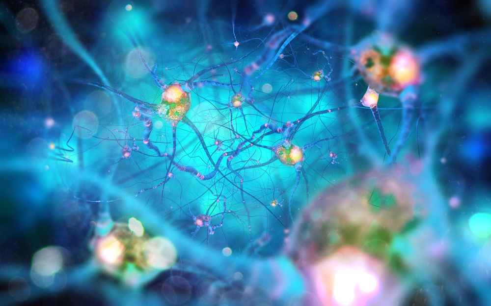
Andrii Vodolazhskyi/Shutterstock
Perisynaptic Schwann cells — a specialized type of cell that helps facilitate communication between neurons and muscle cells — express specific molecular markers, which could make it easier to identify and investigate these cells, a new study has found.
Better understanding these cells’ biology and function could prove useful for understanding diseases where neuron/muscle communication becomes impaired, such as spinal muscular atrophy (SMA) and amyotrophic lateral sclerosis (ALS).
The new findings were published in eLife, in a study titled “Specific labeling of synaptic schwann cells reveals unique cellular and molecular features.”
Glia are cells in the central nervous system that do not conduct electrical currents like neurons. Instead, these cells help guide neurons and provide structural support to synapses, which are the point of contact between two nerve cells that allow them to communicate.
Perisynaptic Schwann cells (PSCs) are a specialized type of glia that are known to be important for the function of synapses at neuromuscular junctions (NMJs), where nerve cells communicate with muscles.
Understanding the function of PSCs may prove invaluable for understanding neuromuscular diseases, such as SMA and ALS.
An obstacle in the understanding of PSCs is that, previously, there were no known molecular markers to differentiate these cells from other types of glia. Molecular markers are molecules (usually proteins) that are expressed only by certain types of cells. Understanding these markers allows researchers to quickly and easily identify cells of interest (by identifying cells expressing the marker).
In the new study, researchers explored potential molecular markers for perisynaptic Schwann cells. The team focused its experiments on calcium-binding protein B (S100β) and neuron-glia antigen-2 (NG2), two proteins that have been identified as markers of other glia types.
The researchers created genetically engineered mice such that cells expressing S100β would also express a red fluorescent protein (dsRed), while cells expressing NG2 would also express a green fluorescent protein (GFP). Under a fluorescent microscope, cells expressing either of these markers can be differentiated by the respective color, while cells expressing both of the markers — and therefore both fluorescent proteins — would appear yellow.
“We found a select subset of glia positive for both S100β-GFP and NG2-dsRed specifically located at the NMJ,” the researchers wrote. “Based on the location and morphology of the cell body and its elaborations, we concluded that PSCs are the only cells expressing both S100β-GFP and NG2-dsRed in skeletal muscles.”
Of note, skeletal muscles are those that are attached to bones.
Using a battery of tests, the researchers showed that the incorporation of the fluorescent molecules didn’t greatly impact the function of the neuromuscular junctions, supporting this mouse model as a tool for studying PSCs. They also showed that the mice could be crossed with a mouse model of ALS, generating ALS mice with labeled PSCs.
“Together, these data strongly indicate that this genetic labeling approach can be deployed to study the synaptic glia of the NMJ in a manner previously not possible in healthy and stressed [diseased] NMJs,” the researchers wrote.
Further experiments indicated that NG2 is expressed only by Schwann cells near neuromuscular junctions (there are other types of Schwann cells besides PSCs), further supporting NG2 as a specific marker of PSCs. The labeled PSCs could be isolated to analyze their transcriptomes (essentially, which genes are “on” or “off” in cells).
With this new model, “it is now possible to determine which cellular and molecular determinants are critical for PSC differentiation, maturation, and function at the NMJ,” the team wrote. “It will also allow us to ascertain the contribution of PSCs to NMJ repair following injury and NMJ degeneration during normal aging and the progression of neuromuscular diseases, such as Amyotrophic Lateral Sclerosis (ALS) and Spinal Muscular Atrophy (SMA).”
“What this means is that we can finally figure out how all three cellular constituents of the synapse — neurons, muscle and glia — talk to each other,” Gregorio Valdez, PhD, study co-author and professor at Brown University, said in a news release.
Although this study focused on the NMJ, the researchers “also gathered initial evidence indicating that synaptic glia cells in the brain can be labeled and targeted using the same approach,” Valdez said. “If true, this discovery could be of immense consequence for treating a myriad of brain conditions, including those involving cognitive decline due to normal aging and Alzheimer’s Disease.”


Leave a comment
Fill in the required fields to post. Your email address will not be published.