
Study reveals front and back of brain develop as two separate organs
Discovery opens new path to study spinal muscular atrophy
Written by |
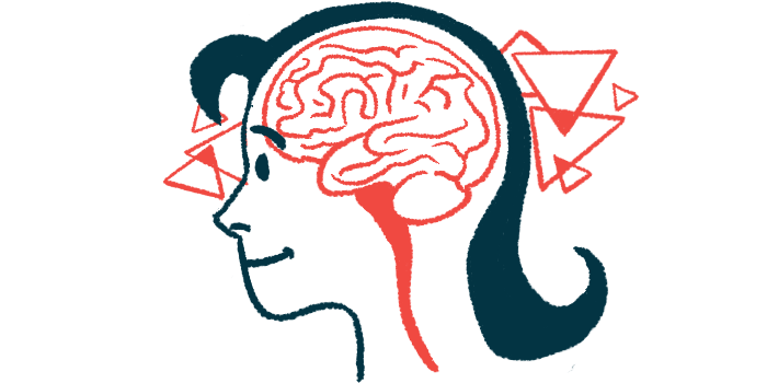
- New research reveals that the front and back parts of the brain develop independently from completely separate progenitor cells.
- This discovery helps explain how SMA affects the hindbrain and allows researchers to grow hindbrain neurons in a lab setting.
- Growing specialized hindbrain progenitor cells provides a critical new cellular model to advance SMA disease research.
The brain has long been thought of as a singular organ, but a new study shows that the front and back parts of the brain actually develop completely independently of each other.
This discovery may have key implications for research into diseases like spinal muscular atrophy (SMA), which affects the back part of the brain but not the front part.
“We’ve shown for the first time that the front of the brain arises from a totally different progenitor cell than the back of the brain,” Kyle Loh, PhD, senior author of the study at Stanford University, said in a university news story. “Our discovery means that we can now grow neurons from the back of the brain, the hindbrain, in a petri dish and study their functions.”
The study, “Two parallel neural ectoderm progenitors contribute to the developing brain,” was published in Nature Neuroscience.
“Scientific advances that give researchers new ways to study the cells and systems affected by neuromuscular diseases are an important part of building a path toward better treatments and, ultimately, improved outcomes for people living with spinal muscular atrophy (SMA),” the Muscular Dystrophy Association (MDA) said in a statement. “While this work is an early research advance and does not represent a treatment … new laboratory models such as these can provide valuable tools for accelerating discovery and deepening our understanding of disease mechanisms.”

Researchers have struggled to grow hindbrain neurons in labs
The adult human brain consists of three main parts: the forebrain, midbrain, and hindbrain. The forebrain and midbrain, which are located closer to the skull, help to control complex thought and emotion. Meanwhile the hindbrain, which is located at the base of the brain just above the spine, is involved in regulating bodily functions and movements.
SMA is a genetic disorder that affects motor neurons, the nerve cells that control movement. The disease likely affects neurons in the hindbrain — but studying how these particular nerve cells are impacted by the disease has proven frustratingly difficult for scientists, because researchers have struggled to grow hindbrain neurons in labs.
The new findings may help explain this long-standing frustration. Scientists have long assumed that all three parts of the brain grow from a single primordial cell during early development. So, when trying to grow hindbrain neurons, scientists have engineered stem cells into brain progenitor cells, then tried to mature them into hindbrain neurons.
“Because specific hindbrain neurons are affected in SMA … and play critical roles in functions including swallowing and breathing, having the ability to study these human cells in the laboratory could help researchers better understand how these diseases affect the nervous system and investigate potential therapeutic approaches,” the MDA said.
Two different primordial cells that give rise to the brain
The new study, done in mice, shows that there are actually two different primordial cells that give rise to the brain: one for the midbrain and forebrain, and a second for the hindbrain. The genetic activity of these two cell types is entirely distinct, and the researchers showed that there’s never any overlap between the two lineages.
“Previous attempts to make hindbrain neurons likely tried to coax forebrain and midbrain progenitors into hindbrain cells, which our study shows is not possible,” said Rayyan Jokhai, a Stanford MD/PhD student and co-author of the study. “In stem cell biology, people are always fixated with creating the end cell type, like the neuron. But it’s important to begin at the earliest stages of embryonic development. Our careful attention to that early time point allowed us to find this fundamental split in brain development.”
Building on this discovery, researchers tried to grow stem cells into hindbrain neurons by first converting the stem cells into the specialized hindbrain progenitor cells — and this was successful. This opens up a new cellular model to study SMA and related neurological diseases.
“Now we have a model to better understand these devastating diseases, and work toward regenerative therapies for them,” Jokhai said. “This is a very exciting new frontier in brain research.”
In addition to opening up new avenues for SMA research, this discovery has key implications for how the brain evolved: the researchers found that the same two-progenitor system is present not only in mice, but also chickens, zebrafish, and even worms that live on the ocean floor.
Our research suggests that evolution took two existing neural systems and pushed them together spatially.
The first animals to evolve nervous systems were jellyfish. These early nervous systems grow at each end of a jellyfish’s body. The researchers’ analyses suggest that, as animals evolved over time and nervous systems became more complex, these two systems became compressed into a single structure that we now call the brain — but from a developmental perspective, there are still two completely separate systems.
“Our research suggests that evolution took two existing neural systems and pushed them together spatially,” Loh said. “Having the brain as one organ would probably be more efficient, but we rely on this primordial way to make the brain as two separate pieces.”
Added Jokhai: “I was surprised at our findings because the word ‘brain’ implies a contiguous organ that likely has a singular origin. But even 500 million years ago, there were these separate neural systems, which now almost operate as one, which is very cool.”


Leave a comment
Fill in the required fields to post. Your email address will not be published.